Near Field Optical Microscope Applications
Nsom Discovering New Worlds Test Measurement Photonics Handbook Photonics Buyers Guide
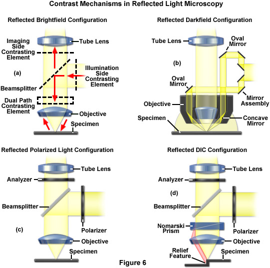
Zeiss Microscopy Online Campus Microscopy Basics Reflected Light Microscopy

Cell Biology Beyond The Diffraction Limit Near Field Scanning Optical Microscopy Journal Of Cell Science
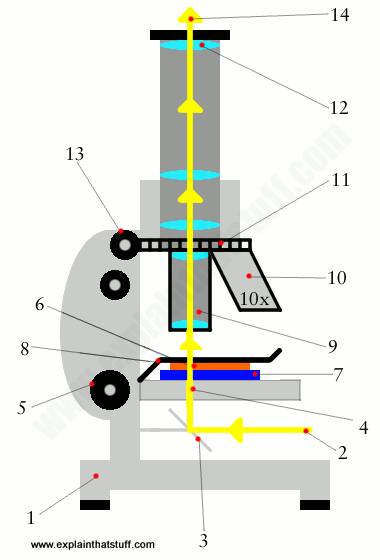
How Does A Microscope Work Explain That Stuff

Lensless Microscopy Chip For Diagnostic Applications Medgadget In 2020 Microscopy Medical Tech Microscope Objective

Nano Antennas Assist In Improving Spatial Resolution Of Terahertz Microscopy Quantum Mechanics Nanotechnology Spatial
Imaging through wavelength refraction and polarization variations snom measurements on algan and uv leds imaging of exciton wave functions time resolved snom measurements.
Near field optical microscope applications. Aperture near field scanning optical microscopy nsom with fluorescence detection gives bio chemical specificity and orientational information in addition to the simultaneously acquired force image. Many different methods have been developed in recent years to gain insight into the structure of proteins membranes organelles and cells. Biological applications of near field optical microscopy article pdf available in ieee engineering in medicine and biology magazine 15 1 51 58 february 1996 with 113 reads. Presents several biological applications of near field optical microscopy in combination with force microscopy.
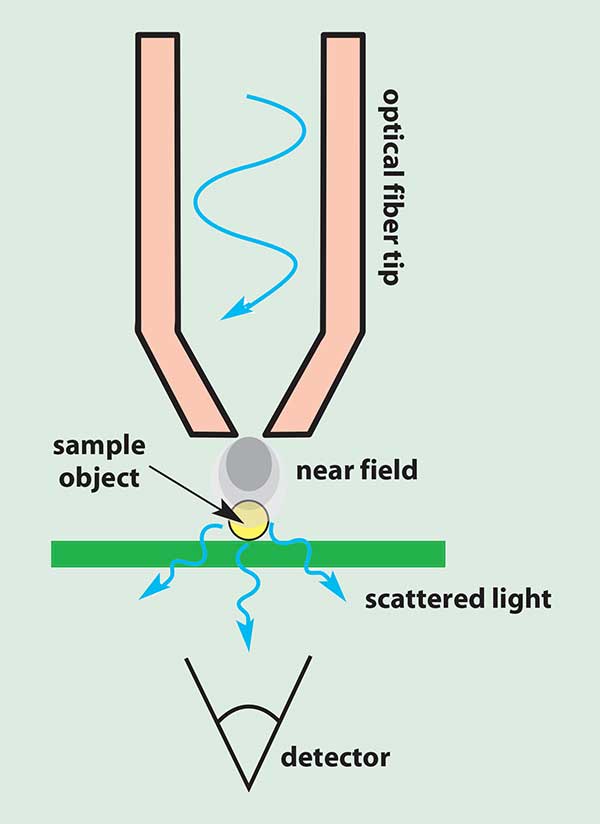
A fundamental principle in diffraction limited optical microscopy requires that the spatial resolution of an image is limited by the wavelength of the incident light and by the numerical apertures of the condenser and objective lens systems. Principle of scanning near field optical microscope and its operation modes. Outline optical near field. Near field scanning optical microscopy nsom or scanning near field optical microscopy snom is a microscopy technique for nanostructure investigation that breaks the far field resolution limit by exploiting the properties of evanescent waves in snom the excitation laser light is focused through an aperture with a diameter smaller than the excitation wavelength resulting in an evanescent.
Scanning near field optical microscopy snom based on metal coated adiabatically tapered fibres combined with shear force feedback and operated in illumination mode has proven to be the most. A new method for high resolution imaging near field scanning optical microscopy nsom has been developed. Probes and distance control. Here we demonstrate the application of near field scanning optical microscopy nsom for analysis of the structures of typical photosynthetic membrane objects such as chloroplasts and thylakoids from spinach and chromatophores from purple bacteria.
The concepts governing this method are discussed and the technical challenges encountered in constructing a working nsom instrument are described.

World S Most Powerful Optical Microscope Lets Researchers See Inside Viruses Popular Science

Nanotech Metasurfaces Will Usher In New Optic Technologies Technology Optical Microscope Physics Research
Zeiss Education In Microscopy And Digital Imaging

Since The Early 1930s Electron Microscopy Has Provided Unprecedented Access To The Alien Wo In 2020 Artificial Intelligence Algorithms Image Patch Systems Engineering
Zeiss Microscopy Online Campus Microscopy Basics Microscope Optical Systems
Molecular Expressions Microscopy Primer Specialized Microscopy Techniques Laser Systems For Optical Microscopy

Optical Microscopy An Overview Sciencedirect Topics
Limitations Of Optical Microscopy
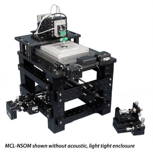
Mcl Nsom Near Field Scanning Optical Microscope

Microscopes An Overview Sciencedirect Topics

Nsom Snom History And Description Nanonics Imaging
Zeiss Microscopy Online Campus Microscopy Basics Enhancing Contrast In Transmitted Light
Instruments Of Microscopy Microbiology

